

How to Choose the Right Fundus Camera for Your Clinic or Distribution Business?
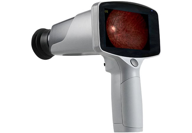
You are sourcing fundus cameras for a new market and the spec sheets all look the same. They are not. The wrong choice at the buying stage creates returns, support headaches, and lost distribution accounts.
Choosing the right fundus camera depends on your clinical setting, patient volume, and whether you need mydriatic or non-mydriatic capability. Entry-level models suit basic diabetic retinopathy screening; mid-range models add autofocus and video for advanced clinics; specialty models extend to veterinary imaging and integrated OCT. Match the model to the actual use case, not the highest spec you can afford.
The fundus camera market rewards specificity. Understand what your buyer actually needs, and the decision becomes straightforward.
What Is the Difference Between Non-Mydriatic and Mydriatic Fundus Cameras?
This is the first question any informed buyer will ask. The answer determines workflow, patient experience, and where the camera can be used.
Non-mydriatic fundus cameras capture retinal images without dilating the pupil. Mydriatic cameras require pupil dilation with eye drops before imaging. Non-mydriatic cameras are faster, more comfortable, and suitable for screening programs where dilation is impractical. Mydriatic cameras offer higher image quality for detailed diagnostic work but require a trained clinician and a longer appointment time.
Clinical Implications of Each Type
Non-mydriatic cameras have become the standard for large-scale screening programs. Diabetic retinopathy screening, for example, is increasingly run through community pharmacies, optometry practices, and mobile units — environments where a trained ophthalmologist is not present and dilation is not practical. A non-mydriatic camera allows a technician to capture usable retinal images in under two minutes per patient.
Mydriatic imaging is still preferred for detailed diagnostic assessment. When an ophthalmologist needs to evaluate the full extent of a retinal lesion, document optic disc changes, or plan treatment for macular degeneration, dilation improves the field of view and image quality significantly.
| Feature | Non-Mydriatic | Mydriatic |
|---|---|---|
| Pupil dilation required | No | Yes (eye drops) |
| Exam time | 1–3 minutes | 20–30 minutes (incl. dilation wait) |
| Image quality | Good (pupil-dependent) | Excellent (wider field) |
| Clinical personnel required | Technician-level | Ophthalmologist preferred |
| Patient comfort | High | Moderate (temporary blurred vision) |
| Best for | Screening programs, high volume | Detailed diagnosis, specialist clinics |
Most modern fundus cameras sold for distribution are non-mydriatic by default. Mydriatic capability is either built-in or available as a mode switch.
How Do Resolution and Field of View Affect Diagnostic Value?
Megapixel count and field of view are the two specifications buyers cite most often. Neither number alone tells the full story.
Resolution determines the fine detail visible in a fundus image — whether small microaneurysms, fine vessel changes, or early optic nerve fiber layer thinning are detectable. Field of view determines how much of the retina is captured in a single image. For diabetic retinopathy and glaucoma screening, wider field of view reduces the number of images needed and increases detection sensitivity.
Resolution: What Matters in Practice
A 12 MP sensor is adequate for most screening applications. For documentation purposes — capturing obvious pathology for referral or patient records — this resolution is more than sufficient. Where resolution becomes critical is in detecting subtle early-stage changes, particularly in monitoring programs where sequential images are compared over months or years.
Higher resolution also matters when images are used for telemedicine referral. The remote reader needs enough detail to make a confident clinical decision from the image alone.
Field of View: Wider Is Not Always Better
Standard fundus cameras offer a 45° field of view. Wide-field cameras extend this to 100° or more. For most primary-care screening purposes, 45° is sufficient. It captures the macula and optic disc — the two areas most relevant to diabetic retinopathy and glaucoma screening.
Wide-field imaging becomes important when evaluating peripheral retinal pathology: lattice degeneration, peripheral breaks, retinal detachment, or conditions like sickle cell retinopathy. For distributors selling into markets with high volumes of these conditions, wide-field capability is a differentiator worth discussing with buyers.
What Models Suit Veterinary Imaging Needs?
Veterinary ophthalmology is a growing segment that many distributors overlook. The equipment needs are specific and the market has less competition than the human clinical segment.
Veterinary fundus imaging requires cameras that can work with the different eye anatomy of dogs, cats, horses, and other animals. Hongdee's FC-161 model is specifically designed for veterinary applications, capturing clear fundus images in small and large animals without requiring the same fixation cooperation that human patients provide.
Key Differences in Veterinary Fundus Imaging
Animals cannot be instructed to fixate on a target. The camera must work quickly, with a wide enough pupil aperture to capture usable images even under challenging conditions. Veterinary patients are often uncooperative, require physical restraint, and may need sedation for detailed examination.
The FC-161's design accounts for these challenges. The imaging protocol is adapted for the shorter examination windows available in veterinary practice. The resulting images support diagnosis of inherited conditions common in specific breeds — progressive retinal atrophy in dogs, for example — as well as trauma and systemic disease affecting the retina.
For distributors, veterinary practices represent a distinct buying audience with different purchasing authority (veterinarians, not ophthalmologists) and different procurement processes (often direct purchase rather than hospital tender). The sales approach needs to be adapted accordingly.
How Do You Price Fundus Cameras for Your Distribution Market?
Pricing strategy for fundus cameras varies significantly by market and by the product tier you are selling.
Entry-level non-mydriatic fundus cameras (like Hongdee's FC-100) are positioned for screening programs and small optometry practices in price-sensitive markets. Mid-range models with autofocus and video capability (FC-200) suit established specialist clinics. Integrated OCT-fundus systems (FC-3100) target hospital ophthalmology departments with larger capital budgets and procurement processes.
Market Segmentation by Price Tier
| Tier | Model Type | Target Buyer | Key Selling Point |
|---|---|---|---|
| Entry | Basic non-mydriatic | Screening programs, new clinics | Low barrier to entry, fast ROI |
| Mid-range | Autofocus, video-capable | Established specialist practices | Efficiency, documentation quality |
| Specialty | Veterinary-specific | Vet hospitals, animal ophthalmology | Niche, lower competition |
| High-end | Integrated OCT+fundus | Hospital departments, academic centers | Single-device comprehensive imaging |
Pricing in emerging markets requires accounting for import duties, local certification costs, and after-sales support infrastructure. A fundus camera that looks competitively priced ex-factory may become expensive once all landed costs are calculated. The distributor margin needs to absorb these costs while remaining competitive against local alternatives.
Hongdee's 12-month warranty, OEM/ODM capability, and 2–5 week lead times are direct margin protectors for distributors. They reduce the cost of returns, enable private-label positioning at higher price points, and allow reliable inventory planning. These are the numbers to include in your business case when evaluating the fundus camera line.
Conclusion
The right fundus camera matches resolution and field of view to the actual clinical need — not the highest specification available. Non-mydriatic models dominate screening markets; mydriatic and wide-field models serve specialist diagnosis. Veterinary imaging is a distinct and growing segment with specialized requirements. Price your distribution offer against landed cost, not just ex-factory pricing.