
Home > Products > Ophthalmic Equipment > OCT
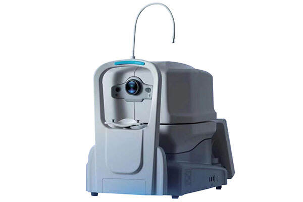
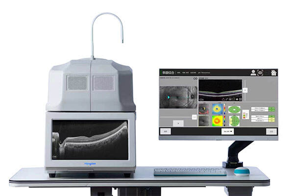
U need precise cross-sectional imaging of retinal layers to diagnose macular degeneration, glaucoma, or diabetic macular edema. Hongdee’s OCT system offers high-resolution B-scans and 3D volume rendering. Its integrated software provides retinal thickness mapping and optic nerve head analysis, ensuring swift decision-making during patient consultations.
- High-speed scanning: Swept-source engine provides rapid volume acquisition.
- User-friendly software: Intuitive interface supports retinal thickness maps, RNFL analysis, macular cube, and glaucoma protocols.
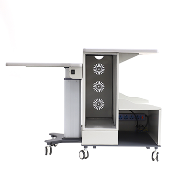
- Compact footprint: Desktop-style unit fits most exam lanes without major reconfiguration.
| Methodology | spectral domain OCT |
| Axial resolution | ≤6 um(in tissue) |
| Transverse resolution | ≤20(in tissue) |
| Scan depth | ≥ 2.5mm(in air) |
| Scan range | 6-12mm |
| Scan speed | ≥ 24,000 A-scan/sec, up to 36,000 A-scan /sec |
| Scan mode | 3D (Macular & Optic Disk), HD,Raster,Circle , Cross |
| Analysis mode | Up to 7 retinal layers segmentation, Macular analysis mode, RNFL & Optic Disk analysis mode, Glaucoma analysis mode and Progress analysis for follow-up examination. |
| Fundus image | OCT en face |
| Focus adjustment | -15D to +15D |
| Pupil diameter | ≥3mm |
| OCT light source | 840nm SLD |
| Optical power | 750um(at cornea) |
| Operation | 13.3inch touch screen ,optional external mouse or keyboard |
| Power supply | 100-240V,50/60 HZ |
| Dimensions | 497 x395x490mm |
| Weight | 34kg |
- Clinical versatility: Supports retina, glaucoma, and anterior segment modules as optional upgrades.
- Seamless workflow: DICOM output integrates with EMR systems for streamlined record-keeping.
- Reliable servicing: Dedicated Hongdee service team ensures calibrated performance and minimal downtime.

We specialize in providing high-quality diagnostic devices, refraction systems, low vision aids, and ophthalmic furniture — all backed by a complete supply chain, strong OEM/ODM service, and a customer-first philosophy.